Backscattered electron images show the differences in the average atomic number of materials within a sample. They are extremely useful in identifying the different phases that make up a sample and their relationships to each other. As the average atomic number of a phase depends on all the all the atoms present in that phase, BSE images reveal the overall chemical differences between the phases; as opposed to x-ray maps which show the differences in concentration of a single element in different phases. In addition, BSE images can usually be acquired much faster than x-ray maps. The two techniques are extremely complimentary in examining the makeup of heterogeneous samples.
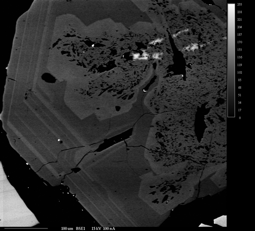
Backscattered electron image of a zoned calcic garnet from a skarn deposit.
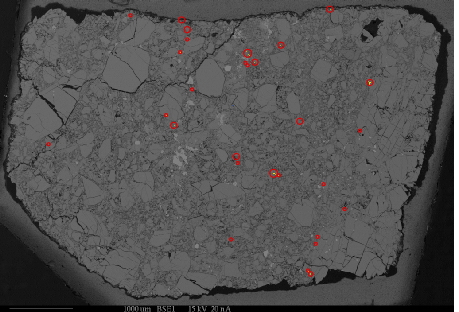
Phosphorus x-ray map overlaid on a backscattered electron image of the same area. Bright areas (highlighted by round, red circles) indicate location of apatite grains. Using this method, grains of the material of interest as small as 2µm can be located for later analysis.